Dental implants can pose difficulties for an individual having an MRI (magnetic resonance imaging), but do not automatically rule out undergoing the procedure, according to Frank G. Shellock, PhD. Shellock has studied the safety and biological effects of magnetic resonance imaging for 25 years and has identified several areas of concern for those with dental implants.
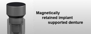
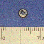
 The use of magnets is popular in dentistry. Following the introduction of powerful cobalt-samarium magnets in the sixties, their use has increased and it is now possible to make them of small dimensions i.e. a few millimetres in width and height. Magnets have found application in two dental specialities namely orthodontics  and prosthetic dentistry . In orthodontics the large attractive and repellent forces of these magnets have been used to correct malocclusions. Conventional magnets have been used as retentive devices for removable partial dentures, obturators and maxillofacial prosthesis. In particular the retentive force and compactness of the rare earth magnets have resulted in their widespread use for overdentures.
Magnetic retention offers many advantages as it serves to dissipate lateral functional forces. There is less need for parallel abutments as a rigid line of insertion is not as critical. Futhermore, the technique is simple, involving minimal time at the chairside and in the laboratory. However careful laboratory handling is required so as not to damage the outer metal coating. Magnets have been used as a retentive element for overdentures. Generally a reversed split pole magnet is used and this is in contact with a ferromagnetic keeper. The technique consists of constructing the keeper in a similar manner to a gold coping. This is cemented into the root face which is then ready for the magnets
Types of Magnets
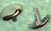
There are two different types of alloys used for the manufacture of small dental magnets. The first alloy used was cobalt-samarium magnets introduced in the sixties. A second alloy based on iron- neodymium-boron became available in the eighties. Both these rare earth magnets have high attractive forces. However, they are brittle materials with a low corrosion resistance. To overcome these problems the magnets are encapsulated in stainless steel, titanium or palladium. If wear of this coating occurs then the alloy will come into contact with saliva. The combination of saliva contact and wear has a deleterious effect on the corrosion resistance of the material and may be increased by the presence of bacteria such as Streptococcus sanguis. This corrosion problem is well documented and can significantly affect the useful life span of intra-oral magnets.
Demagnetization
Dental implants may use magnets to hold the appliance in place. An MRI machine uses powerful magnets that can demagnetize and damage the magnets in the implant, necessitating surgery to repair or replace the appliance. Implants made of ferromagnetic materials use counter-force to keep them in place and appear safe when used in an MRI system operated at 3-Tesla (a measurement of the concentration of a magnetic field) or below.
Many of the dental implants, devices, materials, and objects evaluated for ferromagnetic qualities (an object having a high susceptibility to magnetization, the strength of which depends on that of the applied magnetizing field, and that may persist after removal of the applied field. This is the kind of magnetism displayed by iron.) exhibited measurable deflection forces (e.g., brace bands, brace wires, etc.) but only the ones with magnetically-activated components present potential problems for patients during MR procedures. The issues that exist for magnetically-activated dental implants include possible demagnetization of the magnetic components and the substantial artifacts that these magnets produce on MR imaging.
Reduced Image Quality
Studies of image quality in individuals with dental implants showed 12 of 16 had evidence of deflection of the MRI’s magnetic field that resulted in decreased quality of the image. The MRI may show image artifacts, features that are visible on the image but are not really present.
Patient Injury
The magnetic field may exert sufficient force that a dental implant can dislodge and cause injury to the individual; however, implants properly anchored in surrounding tissue should not cause a problem. The effects of MRI-caused heating of the implant at 3-Tesla is not known.
Under what conditions would a patient with an implant with ferromagnetic components to be to be scanned?
It depends on: the type of implant (electrically active or not), what the implant is made of, and also how long since it was inserted.
If there is no electrical activity associated with the implant and it is non-ferromagnetic (e.g., elgiloy, phynox, MP35N, titanium, titanium alloy, nitinol, tantalum, etc.), the patient can be scanned immediately after implantation. If the implant is weakly magnetic then you should wait six to eight weeks for it to be held firmly in the tissue. (If there is any concern over the ability of the tissue to hold the implant however, don’t scan.)
In general, dental implants, devices, and materials made from ferromagnetic materials tend to be held in place with sufficient counter-forces to prevent them from causing problems related to movement or dislodgment in association with MR systems operating 3-Tesla or less.

