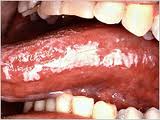
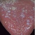
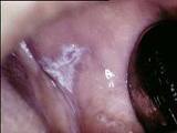
The normal color of soft tissues in your mouth are usually red, with occasional blackish-brownish pigmentation. The difference in color from the rest of your body is due to absence of keratin over the “skin” of your mouth, as evident all over your body. The red color is formed by the blood vessels beneath the thin layer covering in your mouth.
If you find a white patch anywhere in your mouth, it could be a number of conditions or due to a number of reasons as follows:
1. Hereditary lesion
White sponge naevus and leukoedema are examples of lesions that are inherited and are present from young. White sponge naevus, also known as Cannon’s disease or oral epithelial naevus, can be present from infancy or early childhood, and can affect any part of the soft tissues of the mouth. It often occurs bilaterally, meaning on both right and left side of the mouth at the same time. Leukoedema is seen in almost 90% of adult blacks. A positive result with “Stretch test” is a characteristic feature of this lesion, in which the lesion disappears upon stretching or pulling of the involved area.
2. Due to trauma
i) Chemical trauma– the use of aspirins, tobacco products, betel nuts, spices in candies or chewing gums, especially cinnamon and peppermint can cause whitening of soft tissues of the mouth. Those caused by aspirin and tobacco products are termed “aspirin burn” and “tobacco pouch keratosis” respectively.
ii) Thermal trauma– chronic smokers of cigarettes, cigars, and pipes often develop white patches, usually on the inner cheek area, tongue, roof of the mouth or lips. “Pipe smoker’s palate”, or “nicotinic stomatitis” is a phenomenon whereby the roof of the mouth (the palate) appears greyish-white over a large area with scattered red spots all over.
iii) Mechanical trauma- this could be due to cheek biting over long periods of time, a sharp tooth, or prolonged wearing of dentures that do not fit well.
3. Infection-
i) Fungal
Fungal infection, or candidiasis, can be due to a variety of causes such as denture wearing, poor denture hygiene, dry mouth, chronic antibiotic usage, systemic diseases such as anemia, diabetes and acute leukemia. Fungal infection in the mouth can be further divided into:
-pseudomembranous candidiasis, “oral thrush”:
appears as a thick white coating on soft tissue surfaces of the mouth, which can be wiped away leaving a red, raw, and often bleeding surface.
-erythematous or atrophic candidiasis:
it is reddish in color, hence shall not be discusses further in this article
-lesions associated with candida (fungus):
this includes “denture stomatitis”, which is present under areas covered by dentures; “angular chelitis”, which occurs on the corners of the mouth, associated with soreness and some redness; and “median rhomboid glossitis” which as its name suggests, is present on the middle (median) surface of the tongue (“glosso”) and is rhomboid in shape. This latter lesion is also red in color.
-candidiasis associated with systemic conditions as mentioned above.
ii) Bacterial (syphillitic leukoplakia)
Syphillis is an infection caused by the spirochete (a type of bacteria) Treponema pallidum. White patches appear on the tongue in the late (or tertiary) stage of this infection, and carcinoma of the tongue often develops soon after.
iii) viral (hairy leukoplakia)
Hairy leukoplakia presents as a white patch, or often, as vertical white fold, usually occuring on the sides of the tongue, which cannot be removed. It is associated with the Eipstein-Barr Virus (EBV) and is seen largely in HIV-infected patients.
4. Idiopathic (cause unknown)
Any white patch that cannot be characterized clinically or histologically as any other definable disease is classified as “leukoplakia“. Leukoplakia can occure anywhere in the mouth, but in Western Europe and North America, the areas beneath the tongue and the inner cheek areas are considered the most common sites. This lesion should be monitored for any malignant changes such as ulcerations or redness, as they have about 5% chance of turning malignant, ie cancerous.
5. Dermatologic (related to skin diseases)
Some white patches occur concurrently with skin lesions, such as :
i) Lichen planus-
often occurs on the inner cheek region and tongue, but can also involve tongue, roof of the mouth and lips. It usually occurs bilaterally, and is associated with pain and discomfort, which can be aggravated by spicy food.
ii) Lupus erythematosus:
there are 2 types: Discoid lupus erythematosus (DLE) affects only skin, usually face and appears as red patches on cheeks and nose, giving a “butterfly pattern”. Systemic lupus erythematosus (SLE), on the other hand, affects almost all the organs of the body. Lesions in the mouth appear as reddish areas or ulcerations with a whitish border.
6. Tumors and neoplasms:
Eg:Â squamous cell carcinoma

