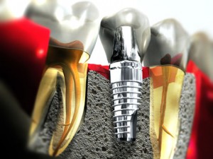
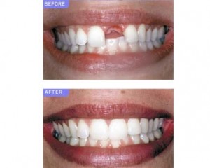
Dental implants are now increasingly used to attach crowns, bridges or dentures by anchorage to bone. Despite its benefits, placement of implants requires careful patient selection and treatment planning to ensure long-term success of implants.
History of dental implants
In 1952, professor Branemark, a Swedish surgeon, while conducting research into the healing patterns of bone tissue, accidentally discovered that pure titanium comes into direct contact with the living bone tissue, the two literally grow together to form a permanent biological adhesion. He named this phenomenon ‘osseointegration’.
Indications for dental implant treatment
- Difficult toothless cases
- Long span dental bridges
- Free end saddles of partial dentures
- Single tooth replacement
Contraindications to tooth implant placement
- Acute illness
- Severe lesions of mucous membranes
- Poor bone quality
- Uncontrolled metabolic disease
- Tumor radiation to implant site
- Unrealistic expectations
- Improper motivation
- Lack of operator experience
- Unable to restore with prostheses
- Cardiac disease – patients with risk of endocarditis may be suitable but regular monitoring is advisable. Contraindicated in severe heart disease
- Hematological disease – contraindicated in hemophilia, relative contraindication for those under Warfarin therapy
- Immunological problems – implant survival may be reduced in patients on corticosteroids and this should be balanced against potential benefits. Smoking has adverse effects on implants
- Bone disorders – most bone disorders are contraindicated. Relative contraindications for osteoporosis
Osseointegation
Osseointegration was defined as ‘a direct structural and functional connection between living bone and the surface of a load carrying implant’. It is in fact an ankylosis of implant to bone surface and some prefer the term functional ankylosis. Osseointegration is prerequisite for success of implant placement.
Factors required for successful osseointegration
- A suitable biocompatible material
- Host bed
- Adaptation of implant to prepared bone site
- Adequate bone quality and quantity
- Atraumatic surgery to minimize tissue damage
- An immobile, undisturbed healing phase
- Soft tissue to implant interface
Suitable biocompatible material
This is necessary to promote healing without a foreign body rejection by host tissue. If biocompatible materials are not used, bone attempts to isolate the foreign body by surrounding it with granulation tissue and then connective tissue. Researchers have demonstrated that titanium and certain calcium-phosphate ceramics are biologically inert.
Host bed
In cortical (outer layer) bone resorption of mineralized, avascular necrotic tissue must occur before new bone can form on the implant surface. In the spongious (inner layer) region of the site, on the other hand, woven bone formation and osseointegration occurs early in the process of healing.
Adaptation of implant to prepared bone site
Site of gap between the implant and the bone immediately after implant placement is critical to achieving osseointegration. Gap size can be controlled primarily by the preparation of a precise surgical bed. Precision instrumentation and technically sound surgical procedure minimize the distance between the implant and host bone.
Atraumatic surgery to minimize tissue trauma
Atraumatic surgery is required to allow minimal and thermal injury to occur.
Adequate bone quality and quantity
Implant immobility during the healing phase is affected by bone quality and quantity. Areas with high cortical (outer layer) bone such as the front of the lower jaw are more likely to anchor the implant successfully. Initial stability takes long time to achieve in cancellous (porous) bone. Implant stability increases if both superior and inferior cortical plates are used.
Upper jaw
- cortical plate of upper jaw is often thin or absent and normally crumbles,
- pattern of bone resorption  and anatomical structures in upper jaw cause problems,
- resorption causes bone loss from front and crestal surfaces
Lower jaw
- more cortical bone so better success rate,
- resorption in lower jaw does not pose problem with implant placement but there is severe loss of bone height
 Methods of improving implant success in the upper jaw
- Use additional implants to share the load
- Use connecting bars
- Use maximum length of implant
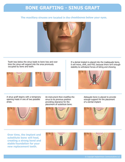
- Consider augmentation of ridge bone
- Consider sinus lift to extend available ridge
- Allow more time for osseointegration
Undisturbed healing phase
It is fundamental that undue loading is avoided until osseointegration is achieved.
Minimum integration time
Region of implant placement |
Minimum integration time |
|
Front of the lower jaw |
3 months |
|
Back of the lower jaw |
4 months |
|
Front of the upper jaw |
6 months |
|
Back of the upper jaw |
6 months |
|
Into bone graft |
6 to 9 months |
Soft tissue to implant interface
Successful dental implant should have an unbroken, perimucosal seal between soft tissues and implant abutment surface. To maintain the integrity of this seal, the individual must maintain a high level of oral hygiene. If the seal between soft tissue and implant is lost, the gum pocket can extend directly to bony structures. Therefore if the seal breaks down or is not present, the area is subject to peri-implant gum disease – peri-implantitis
To be continued in Part 2


