Continued from Part 1
Cold testing
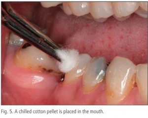
A cold stimulus such as refrigerant spray is sprayed on a cotton pledget and applied to the tooth under investigation.
Heat testing
A heated source such as a GP stick is applied to the tooth.
Test cavity preparation
In extreme cases, a cavity is prepared in a tooth under copious irrigation with no local anesthetic, using a sharp bur as a last resort. Indiscriminate test cavity preparation is not done particularly when teeth are restored with expensive restorations such as crowns and bridges.
- If there are no signs and symptoms with tooth and there is a normal response to sensory stimuli, the pulp is normal or healthy.
- If decay is close to the pulp but has not penetrated it, the condition is termed reversible pulpitis. The endodontist may place a sedative material in the tooth and wait to determine if the tooth will repair itself.
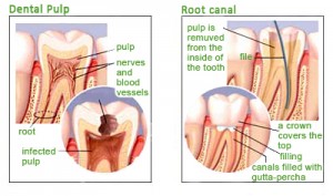
- If bacteria exist in the pulp, the condition is called irreversible pulpitis and root canal treatment is necessary.
- If tooth does not respond to sensory stimulus, the pulp is deemed necrotic or dead.
Radiographic examination
All aspects of root canal practice are heavily reliant upon information gains from radiographs or x-rays. X-rays are taken pre-operatively, during treatment and post-operatively.
Root canal procedure
Root canal treatment (RCT) involves the removal of infected root canal and pulp tissues as well as cleaning and filling of the resultant space, in order to prevent microorganisms from multiplying within the root canal system and spreading to the gum tissues. Root canal therapy usually requires around three to four appointments and no dental procedure is without risks. Pain, surgery and the chance of an infection developing may happen during root canal treatment.
Anesthesia and pain control
- Local anesthetic agents are injected to numb the tooth.
Isolation and disinfection of operating field
- A rubber dam will be placed on the tooth concerned to keep bacteria and saliva at bay.
- Antiseptic solution is swabbed over exposed tooth, the clamp and the surrounding dental dam.
Accessing the tooth and removal of pulp
- The endodontist enters the crown portion of the tooth with a tungsten carbide bur, removing decay and infected tooth structure.
- Once the root canals are located with the endodontic explorer, the pulp tissues are removed with a brooch.
- The canals are irrigated gently with the sodium hypochloride solution and saline solution.
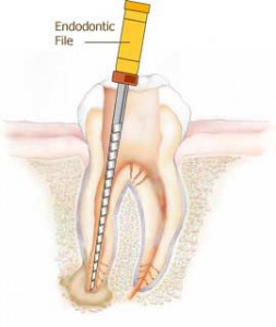
- The endodontist uses a small endodontic file to rub the irrigation solution against the walls of the canal and pulp chamber.
Cleaning and shaping the canal
- The endodontist inserts files into the canal and moves them up and down with short strokes. During this motion, the cutting edges of the files remove dentin and debris from the walls.
- An x-ray of the tooth with a trial file in the canal. This is the working length radiograph.
- The root canals are cleaned and shaped to enlarge the diameter of the canals.
- The canals are irrigated thoroughly at frequent intervals during this shaping and cleaning process to prevent dentin shavings from clogging the cutting edges.
- Paper points are used for insertion into the canals until the points come out dry.
Preparing to fill the canal
- An appropriate-sized gutta-percha point is selected, and cut it to the predetermined length. This is called the master cone.
- An x-ray of the tooth with the master cone in the canal.
- If the x-ray does not show the tip of the point within 1mm of the apex of the root, the point is repositioned and another x-ray is taken.
Filling the canal
- Once the master cones are snugly fitted into the root canals, the canals are ready to be filled with gutta-percha points.
- A root canal sealer is placed and an endodontic plugger is used to compact the points vertically.
- This routine continues until the canal is completely filled
- The endodontist then places a temporary restoration on the canal.
- A post treatment x-ray is exposed.
- The endodontist checks the bite of the temporary restoration and adjust as needed.
Post treatment instructions and follow-up
- If there are indications of a problem, such as swelling or pain, you should go back to the practice immediately.
- Another visit is necessary to have a final restoration placed. Depending upon the amount of tooth structure left, a simple restoration or a post and core with a crown would be needed. Complex restorations would require further visits.
- A follow-up around 3 to 6 months later with the endodontist should be done to ensure the root canal treatment was a success and that no complications have developed.


