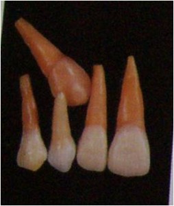
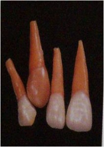
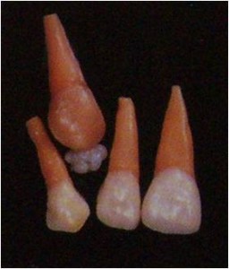
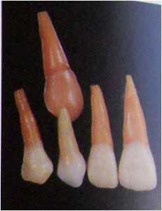
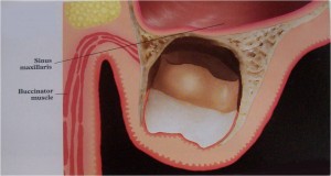
What is Impacted Tooth?Â
An impacted tooth is any tooth that is prevented from reaching its normal position in the mouth by tissue, bone, or another tooth.
Why do impactions occur?
Theories & mechanisms of tooth impaction
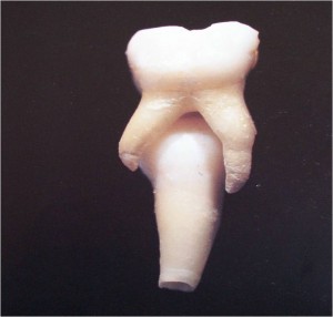
Theories of impaction
By Durbeck
1) Orthodontic theory : Jaws develop in downward and forward direction. Growth of the jaw and movement of teeth occurs in forward direction,so any thing that interfere with such moment will cause an impaction (small jaw-decreased space).
A dense bone decreases the movement of the teeth in forward direction.
2) Phylogenic theory: Nature tries to eliminate the disused organs i.e., use makes the organ develop better, disuse causes slow regression of organ.
[More-functional masticatory force – better the development of the jaw]
Due to changing nutritional habits of our civilization, use of large powerful jaws have been practically eliminated. Thus, over centuries the mandible and maxilla decreased in size leaving insufficient room for third molars.
3) Mendelian theory:Heredity is most common cause. The hereditary transmission of small jaws and large teeth from parents to siblings. This may be important etiological factor in the occurrence of impaction.
4) Pathological theory: Chronic infections affecting an individual may bring the condensation of osseous tissue further preventing the growth and development of the jaws.
5) Endocrinal theory: Increase or decrease in growth hormone secretion may affect the size of the jaws
What will happen if impact teeth are retained?
Complications

Infections:
Pericoronal infection
Acute / chronic alveolar abscesses
Chronic suppurative osteitis
Necrosis
Osteomyelitis
Complications
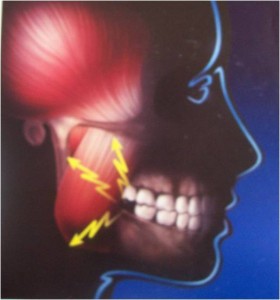
Pain:
Slight and restricted
Severe or excruciating
Intermittent, constant or periodic
Referred to ear, the post auricular area, any part of the area supplied by the trigeminal nerve. (Eg. Temporal pain)
Fractures:
Impacted tooth proves that weakening of the mandible occurs due to displacement of bone.
Other complications:
Ringing, singing or buzzing sound in the ear (Tinnitus aurium)
Otitis
Affections of the eye such as
Dimness of the vision
Blindness
Iritis
Pain simulating that of glaucoma
Indications and contraindications for removal of impacted tooth
“A strong indication for removal of impacted third molar should be complemented with a strong contraindication to its retentionâ€
– Mercier P., Precious D., Risk and benefits of removal of impacted third molars, IJOMS 21:17, 1992.
Indications:
Pericoronitis – 27% to 34% (Swed Den J1987)
Caries – 3% to 15% (IJOMS 1988)
Root resorption – 5% (Swed Den J 1987)
Formation of follicular cyst – 1 to 5%(J Oral Pathol 1998)
Tumors arising in the follicular (Dentigerous cysts) – 0.1 to 0.2% (JOMS 1991)
Contraindications:
Acute infection with pericoronitis
Medically compromised state – uncontrolled diabetes
Extremes of age – Old age
Basic principles of impactions
1. Any region of the dental follicle proper has the potential for initiating and regulating bone resorption and bone formation or not influencing bone activity.
Ectopic eruption is easily explained by aberrant activation of the follicle.
2. Movement of teeth during eruption consists of preparing a path through bone / soft tissues and moving them along this path.
3. Formation of the eruption pathway is the rate-limiting step as revealed by rapid catch-up eruption of temporary unerrupted teeth.
Causes for Eruption Disturbances
Rationale treatment approach
It is therefore necessary to develop a rationale treatment approach in order to diagnose which of the prerequisites for eruption have been violated and to what extent.
This diagnosis encompasses a
History
Clinical examination
Detailed radiographic examination allowing a three dimensional concept of the region involved.
Impacted Maxillary Third Molars
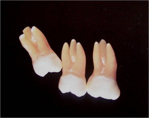
Natural Development
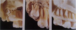
Note the initial position close to the cervical region of the second molar and the gradual uprighting of the tooth germ
Surgical Anatomy
Classification of Maxillary Third Molars
Based on anatomic position ,follows
Relative depth of the impacted maxillary third molar in bone
The position of the long axis of the impacted maxillary third molar in relation to the long axis of the second molar
Relationship of the impacted maxillary third molar to the maxillary sinus
Relative depth of the impacted maxillary third molar in bone:
Class A: The lowest portion of the crown of the impacted maxillary third molar is on a line with the occlusal plane of the second molar
Class B: The lowest portion of the crown of the impacted maxillary third molar is between the occlusal plane of the second molar and the cervical line.
Class C: The lowest portion of the crown of the impacted maxillary third molar is at or above the cervical line of the second molar.
