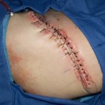
Wound closure techniques have evolved from the earliest development of suturing materials to comprise resources that include synthetic sutures, absorbables, staples, tapes, and adhesive compounds. The engineering of sutures in synthetic material along with standardization of traditional materials (eg, catgut, silk) has made for superior aesthetic results. Similarly, the creation of natural glues, surgical staples, and tapes to substitute for sutures has supplemented the armamentarium of wound closure techniques. Aesthetic closure is based on knowledge of healing mechanisms and skin anatomy, as well as an appreciation of suture material and closure technique. Choosing the proper materials and wound closure technique ensures optimal healing.
Wound healing
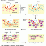
 Three phases of wound healing have been identified and studied on the cellular and molecular level. These 3 distinct phases, ie, inflammation, tissue formation, and tissue remodeling, depend on an elaborate cascade of growth factors and cellular components interacting in a directed manner to achieve wound closure.
The initial injury leads to the recruitment of inflammatory cells into the wound, once a clot forms in response to disrupted blood vessels. This scenario entails a complex interaction between local tissue mediators and cells that migrate into the wound. The inflammatory phase occurs in the first few days as inflammatory cells migrate into the wound. Migration of epithelial cells has been shown to occur within the first 12-24 hours, but further new tissue formation occurs over the next 10-14 days.
Epithelialization and neovascularization result from the increase in cellular activity. Stromal elements in the form of extracellular matrix materials are secreted and organized. This new tissue, called granulation tissue, depends on specific growth factors for further organization to occur in the completion of the healing process. This physiologic process occurs over several weeks to months in a healthy individual.
Finally, tissue remodeling, in which wound contraction and tensile strength is achieved, occurs in the next 6-12 months. Systemic illness and local factors can affect wound healing. Traditionally, at least 2 types of wound healing have been described, ie,
primary intention and secondary intention.
In the primary intention method, surgical wound closure facilitates the biological event of healing by joining the wound edges. Surgical wound closure directly apposes the tissue layers, which serves to minimize new tissue formation within the wound. However, remodeling of the wound does occur, and tensile strength is achieved between the newly apposed edges. Closure can serve both functional and aesthetic purposes. These purposes include elimination of dead space by approximating the subcutaneous tissues, minimization of scar formation by careful epidermal alignment, and avoidance of a depressed scar by precise eversion of skin edges. If dead space is limited with opposed wound edges, then new tissue has limited room for growth. Correspondingly, atraumatic handling of tissues combined with avoidance of tight closures and undue tension contribute to a better result.
The secondary intention method (spontaneous healing) is ancient and well established. It can be used in lieu of complicated reconstruction for certain surgical defects. This method also depends on the 3 stages of wound healing to achieve the ultimate result.
Adhesives
Use of surgical adhesives can simplify skin closure in that certain problems inherent to suture use can be avoided. Problems (eg, reactivity, premature reabsorption) can occur with sutures and lead to an undesirable result, both cosmetically and functionally.
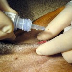
Low tension wounds (those where the skin edges lie close together without significant tension) can be closed by gluing the skin edges together with a skin adhesive.Several adhesives have been developed to alleviate this problem and to facilitate wound closure. One substance, cyanoacrylate, has been used for 25 years and easily forms a strong flexible bond. In some forms,
it can induce a substantial inflammatory reaction if implanted subcutaneously. If used superficially on the epidermal surface, little problem with inflammation occurs. In a study on the use of adhesives in the emergency department, adhesives were more likely to be used in facial lacerations and in children and less likely to be used in longer scars. The concomitant use of either a topical anesthetic or no anesthetic, as opposed to an injectable, was cited as an advantage in the use of adhesives.
Octyl-2-cyanoacrylate (Dermabond, Ethicon, Somerville, NJ) is the only cyanoacrylate tissue adhesive approved by the U.S. Food and Drug Administration (FDA) for superficial skin closure. Octyl-2-cyanoacrylate should only be used for superficial skin closure and should not be implanted subcutaneously. Subcutaneous sutures are used to take the tension off the skin edges prior to applying the octyl-2-cyanoacrylate. Subcutaneous suture placement aids in everting the skin edges and minimizing the chances of deposition of cyanoacrylate into the subcutaneous tissues.

