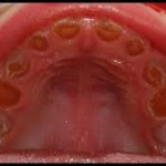
Osteogenesis imperfecta (OI) is always associated with bone fragility. In addition, OI may affect the growth of the jaws and may or may not affect the teeth. About half of the people who have OI have teeth that appear normal, and their major concerns are routine care. However, the other half has a defect in the teeth called dentinogenesis imperfecta (DI), sometimes referred to as opalescent teeth or brittle teeth. These teeth may be misshapen, may chip or break easily, and will require special care.

Osteogenesis imperfecta with dentinogenesis imperfecta
Oral cavity problems related to osteogenesis imperfecta may include the following:
• A skeletal Class III malocclusion. The teeth do not correctly match up making biting difficult. This is caused by the size and/or position of the upper jaw or the lower jaw.
• An open bite. There is a vertical gap between some of the upper and lower teeth.
• Impacted teeth. The first or second permanent molars do not erupt, or they erupt out of the usual location (ectopic).
• Dental development. Tooth development may be delayed or advanced in some individuals affected by OI.
OI does not affect the presence or absence of gum disease (periodontitis).
Major Parts of the Teeth
The teeth are made up of four distinct parts.
• Enamel is the outside part of the crown. It is the hardest substance in the body and the point of contact for chewing.
• Dentin is the substance under the enamel that forms the rest of the crown and surrounding the pulp chamber and almost all of the root structure. It is similar to bone.
• The Pulp Chamber is the inner hollow part of the tooth containing blood vessels and nerves.
• The Dentinoenamel Junction (DEJ) is the term for where the enamel and dentin are attached to each other.
Dentinogenesis Imperfecta (DI)
Dentinogenesis imperfecta can be part of osteogenesis imperfecta (DI type I) or it can be a separate inherited dominant trait without OI (DI type II). DI occurring with OI seems to run in families but can vary in severity from one member to another. DI has a variable affect on the color, shape, and wear of both primary and permanent teeth. If someone has OI and DI, all of their teeth may not be affected to the same degree.
Teeth affected by DI have essentially normal enamel, but the DEJ and the dentin are not normal. The enamel tends to crack away from the dentin, which will wear away more quickly than enamel. The dentin makes the teeth look darker or opalescent. The dentin also grows to fill in the pulp chamber, causing a loss of feeling in the tooth. Affected teeth will have an increased incidence of fracture, wear and decay.
Dentinogenesis imperfecta may be diagnosed with the first baby tooth. If the tooth looks gray, bluish, or brown, DI should be suspected. Children should be taken to a dentist (if possible a specialist in pediatric dentistry) when the first teeth are erupting. This may happen as early as 6 months to 1 year of age. Radiographs, or X-rays, can be useful but may be difficult to obtain until the child is older. Sometimes there are changes visible on the X-rays that are not obvious just by looking at the teeth. Crowns appear bulbous and roots may be shorter and more slender than standard. Primary teeth are usually more affected than the permanent teeth.
When, for any reason, crowns are not feasible, a “tooth color†dental material may be used, such as composites or glass ionomers in conjunction with composites. The sand abrasion method may also be useful because it removes carious dentine only and thus spares dental tissue. In any case, amalgam restorations should not be used because they impose an additional stress on the teeth.
General Care for People With OI and Without DI
A dentist should see a child with OI by 6 months after the eruption of the first baby tooth at the latest. Baby teeth require care. They are important for chewing, speaking, holding space for the permanent teeth to grow in, and growth of the jaws. There appears to be minimal risk of jaw fracture from routine dental care and dental extractions. No particular precautions are needed other than those that would be taken anyway, such as support of a very thin lower jaw when an extraction procedure is being done.
Good care involves brushing and flossing the teeth of young children, then teaching them how to do it themselves and checking them as they grow older. Soft toothbrushes are good for everybody and easier on gums, since gums also need brushing. Mechanical toothbrushes tend to be more effective than brushing by hand. Use of fluoride toothpaste is recommended. Children should use a small dab of toothpaste, or a children’s toothpaste, and be taught to spit it out well after brushing so they do not swallow excessive amounts. Before going to bed, children should spit out the toothpaste after brushing, but not rinse their mouths. This will leave more fluoride in the mouth to work overnight. Parents should talk to their child’s dentist about the fluoride content of their drinking water and ask if supplemental fluoride is needed from a pill, a non-alcohol fluoride rinse, or a fluoride gel. Sealants placed on the biting surface of the permanent molars in children may reduce the chance of developing cavities in the grooves of the teeth.
Starting when the child is 7 years old, an orthodontist should check the child’s bite for evidence of an open bite or Class III malocclusion.


requir dental care advise for 7yers baby of OI type IV.
Thank you for taking time to read my articles. I will try to find some information for you.
Pls check back for the information you want in a week’s time.
thank you