Category Archives: Oral medicine and pathology

Ehler-Danlos Syndrome Part 2

Signs and symptoms

Signs vary widely based on which type of EDS the patient has. In each case, however, the signs are ultimately due to faulty or reduced amounts of collagen. EDS most typically affects the joints, skin, and blood vessels. Following is a list of major signs and symptoms. Continue reading

Oral Pemphigoid

Oral pemphigoid (pemphigoid of the mouth) is an uncommon blistering condition which affects primarily the lining of the mouth and gums. Other moist surfaces of the body (known as mucous membranes) can also be affected, and these include the surface layers of the eyes, inside the nose and the genitalia. The skin is less commonly involved but can be affected by a similar blistering condition, known as bullous pemphigoid. Continue reading

Osteonecrosis of the Jaw Part 2

A brief introduction has been done in “Osteonecrosis of the jaw Part 1“. Here in this article we will be discussing briefly about the prevention and treatment of osteonecrosis of the jaw.

Osteonecrosis-exposed bone on the right edentulous region of the lower jaw

How is osteonecrosis of the jaw treated?

Osteonecrosis can either be treated conservatively, or surgically:

i) Conservative treatment

Conservative treatment basically means that no active treatment is done that is directly addressing the problem. Usually, patients who present with osteonecrosis of the jaw are started on antibacterial rinses (eg: Chlorhexidine gluconate mouthwash), antibiotics and oral analgesics. Continue reading

Osteonecrosis of the Jaw Part 1

In this article we will be discussing questions about osteonecrosis of the jaw such as :

What is it?

How does it happen?

What are the causes & risks for osteonecrosis of the jaw?

What are bisphosphonates?

Should I be concerned if I am on bisphosphonates?

Why is osteonecrosis of the jaw dangerous?

The treatment and prevention of osteonecrosis of the jaw will be further discussed in the article “Osteonecrosis of the Jaw Part 2”.
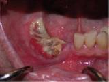
Osteonecrosis- exposed bone that does not heal in the right edentulous region of the lower jaw.

What is osteonecrosis of the jaw?

“Osteonecrosis” is made up of the words “osteo”, which means bone, and “necrosis” which means death of cells. Hence “osteonecrosis” of the jaw bone means death of cells in the jaw bones. It is diagnosed when an area of bone is exposed and shows no sign of healing or gum growing over it 8 weeks after an invasive dental procedure, such as tooth extraction or implant surgeries. Both the upper and lower jaw may be affected, and it may be associated with pain, numbness, swelling and infection of the affected site. The damage to the jaw bone is irreversible, and if left untreated, can spread and can cause devastating damages, even to the point of death. Continue reading

Dry Mouth Mouthwash

Dry mouth, also called xerostomia, is a condition that can interfere with everyday activities, such as eating, talking or sleeping. Some common symptoms of dry mouth include:

  • Bad breath
  • A sticky, dry or sore mouth
  • Cracking at the corners of the mouth
  • A red and parched mouth
  • Blisters and mouth ulcers
  • A pebbled look to the tongue
  • Difficulty eating dry or spicy foods
  • Waking up with a dry mouth at night Continue reading

An Approach to Oral Ulcers

© buzzle.com

Oral ulceration is probably the commonest oral mucosal disease seen and it may also be the most serious. It is important therefore not to underestimate that ulcer in your mouth, especially if it persists for a long time. Continue reading

Odontogenic myxoma

The odontogenic myxoma is an uncommon benign odontogenic tumor arising from embryonic connective tissue associated with tooth formation. As a myxoma, this tumor consists mainly of spindle shaped cells and scattered collagen fibers distributed through a loose, mucoid material. Continue reading

Calcifying epithelial odontogenic tumor

The calcifying epithelial odontogenic tumor was first described by Pindborg in 1956; hence also called Pindborg’s tumor. The calcifying epithelial odontogenic tumor is a benign odontogenic tumor of epithelial origin that accounts for approximately 1% of all odontogenic tumors. The origin of this neoplasm is not clearly known, although it is generally accepted to be derived from oral epithelium, reduced enamel epithelium, stratum intermedium or dental lamina remnants. It is more common in the posterior part of the mandible of adults in the fourth to fifth decades. There is no gender predilection. It is characterized by squamous epithelial cells, calcifying masses, and homogeneous acellular material admixed with the tumor epithelium and stroma that have been identified as amyloid. Continue reading

Ameloblastoma

Ameloblastoma (from the early English word amel, meaning enamel + the Greek word blastos, meaning germ) is a rare, benign tumor of odontogenic epithelium (ameloblasts, or outside portion, of the teeth during development) much more commonly appearing in the lower jaw than the upper jaw. It was recognized in 1827 by Cusack. This type of odontogenic neoplasm was designated as an adamantinoma in 1885 by the French physician Louis-Charles Malassez. It was finally renamed to the modern name ameloblastoma in 1930 by Ivey and Churchill. Continue reading

Anatomy of temporomandibular joint Part 3

The disc is thick all around its rim, and thin in the centre. From anterior to posterior it shows – anterior extension, thick anterior band (2.0mm thick), intermediate thin zone (1.0 mm thick), thick posterior band (3.0 mm thick) and posterior most bilaminar region (Dubrul, 1996; Williams et al, 1999). The disc is attached all around the joint capsule except the strong straps those fix the disc directly to the medial and lateral condylar poles which ensures that the disc and condyle move together in protraction and retraction (Choukas and Sicher, 1960; Williams et al, 1999). The anterior extension of the disc is attached to fibrous capsule superiorly and inferiorly and through that to temporal bone and the mandibular neck respectively. In between it gives insertion to lateral pterygoid muscle where the fibrous capsule is lacking and synovial membrane is supported only by loose areolar tissue. In the opinion of Kreutziger and Mahan (1975), this deficiency anteriorly is the weak point since there is no fibrous resistance to hypertranslation. Apart from lateral pterygoid, anteromedially, there are attached some fibres of masseter and temporalis laterally. Although more than one muscle is inserted into the disc, majority of the interest has been focussed on lateral pterygoid, whose deep position, unfortunately makes it difficult to investigate under natural conditions (Moore). Continue reading