Gardner syndrome which was ï¬rst described in 1953 consists of adenomatous polyps of the gastrointestinal tract, desmoid tumours, osteomas, epidermoid cysts, lipomas, dental abnormalities and periampullary carcinomas.The incidence of the syndrome is 1:14,025 with an equal sex distribution. It is determined by the autosomal dominant familial polyposis coli gene (APC) on chromosome 5. Continue reading
Category Archives: Oral Surgery
Flaps for Facial Reconstruction
Facial flaps can be divided into two types: Axial and Random. An axial flap has a named artery supplying it. The surviving length of an axial flap will remain constant regardless of the width of the flap. A random flap has smaller unnamed vessels and is not as stable. It’s surviving length is in direct proportion to the width. A random flap’s surviving length can be lengthened by “delaying” the flap. To delay a flap, it is elevated but left in position as a bipedicle flap. Two weeks later it is raised as a unipedicle flap and placed into position to close the defect. Interpolation flaps traverse skin in order to reach the defect. If placed over the skin, they will have a pedicle. The pedicle can be divided in 3 to 6 weeks depending upon the type of flap and the condition of the patient. Flaps may be “trained” by occluding the blood supply in the pedicle for progressive lengths of time. This allows for an earlier transection of the pedicle. Continue reading
Oro-antral fistula
This is a fistulous communication between the floor of the maxillary sinus to the oral cavity. This commonly occurs following dental extraction of infected upper molar and premolar tooth. From a small cavity at birth, the maxillary sinus starts to enlarge during the third month of foetal life and usually reaches maximum development around the eighteenth year. Its volume is approximately 20-25 ml in a normal adult. The floor of the sinus consists of the alveolar process and the hard palate. Continue reading
Kaposi’s sarcoma Part 1
Kaposi’s sarcoma (KS) is a tumor caused by Human herpesvirus 8 (HHV8), also known as Kaposi’s sarcoma-associated herpesvirus (KSHV). It was originally described by Moritz Kaposi (KA-po-she), a Hungarian dermatologist practicing at the University of Vienna in 1872. It became more widely known as one of the AIDS defining illnesses in the 1980s. The viral cause for this cancer was discovered in 1994. Although KS is now well-established to be caused by a virus infection, there is widespread lack of awareness of this even among persons at risk for KSHV/HHV-8 infection. Continue reading
Surgical exposure of impacted tooth
When a tooth fails to emerge through the gums, it is considered to be an impacted tooth. This commonly occurs in the case of canine teeth.
It is important to treat an impacted tooth in order to prevent the improper eruption of nearby teeth, cyst formation, possible infection or other negative changes in the jaw. Continue reading
Cleft lip repair Part 2
Surgical procedure
Cleft lip repair can be initiated at any age, but optimal results occur when the first operation is performed between two and six months of age. Surgery is usually scheduled during the third month of life.
While the patient is under general anesthesia, the anatomical landmarks and incisions are carefully demarcated with methylene blue ink. An endotracheal tube prevents aspiration of blood. The surgical field is injected with a local anesthestic to provide further numbing and blood vessel constriction (to limit bleeding). Myringotomy (incisions in one or both eardrums) is performed, and myringotomy tubes are inserted to permit fluid drainage. Continue reading
Osteonecrosis of the Jaw Part 2
A brief introduction has been done in “Osteonecrosis of the jaw Part 1“. Here in this article we will be discussing briefly about the prevention and treatment of osteonecrosis of the jaw.
How is osteonecrosis of the jaw treated?
Osteonecrosis can either be treated conservatively, or surgically:
i) Conservative treatment
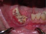
Conservative treatment basically means that no active treatment is done that is directly addressing the problem. Usually, patients who present with osteonecrosis of the jaw are started on antibacterial rinses (eg: Chlorhexidine gluconate mouthwash), antibiotics and oral analgesics. Continue reading
Cleft lip repair Part 1
Cleft lip repair (cheiloplasty) is surgical procedure to correct a groove-like defect in the lip.
Purpose
A cleft lip does not join together (fuse) properly during embryonic development. Surgical repair corrects the defect, preventing future problems with breathing, speaking, and eating, and improving the person’s physical appearance. Continue reading
Fracture of the Lower Jaw-Part 2
We have discussed the principles of management of fractures in Fracture of the Lower Jaw-Part I. In this article we will be discussing a little in depth regarding the management of Lower jaw fractures, or mandibular fractures.
 PRINCIPLES OF MANAGEMENT OF FRACTURES:
- Reduction of the fracture-Reduction can be done in 2 days: Open Reduction or Closed Reduction, as will be discussed below
- Fixation & stabilization of the fracture-Direct or Indirect
- Immobilization of segments at fracture site
- Occlusion restored-to allow the patient to bite in his original position
- Infection eradicated/prevented-infection can prevent or delay healing, thus it is of essence that infection be avoided. Continue reading
Fracture of the Lower Jaw-Part I
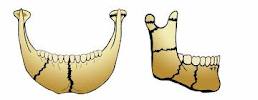
Trauma exerted onto the head and neck region can cause a fracture to any of the bones. The lower jaw, or the mandible, is particularly prone to fracture. In this article we will discuss some of the aspects related to fracture of the lower jaw (mandibular fracture).
CAUSES OF JAW FRACTURE (upper or lower):
- Accidents: Motor-vehicle accidents (MVA), sports injuries, occupational (accidents that occur during work)
- Falls (eg, falling down the stairs, slipping on a slippery floor)
- Assault and fights
- Pathological– a pathology such as tumours or cysts in the jaw bone can cause thinning of the bone or decrease in density of the bone, ultimately leading to fracture of bone in that region, even when a light external force is applied to the bone. Continue reading
