Caries diagnosis Part 1

1. What are the best methods for detecting early and advanced dental caries (validity and feasibility of traditional methods; validity and feasibility of emerging methods)?

Observations and studies during the past two decades have indicated that diagnostic and treatment paradigms may differ significantly for large, cavitated lesions versus early, small lesions and demineralized areas on tooth surfaces. The essential anatomic-pathophysiologic problem is that the carious lesion occurs within a small, highly mineralized structure following penetration through the structure’s surface in a manner which may be difficult to detect using current methods. Additionally, carious lesions occur in a variety of anatomic locations, often adjacent to existing restorations, and have unique aspects of configuration and rate of spread. These differences make it unlikely that any one diagnostic modality will have adequate sensitivity and specificity of detection for all sites. The application of multiple diagnostic tests to the individual patient increases the overall efficacy of caries diagnosis. Existing diagnostic modalities require stronger validation, and new modalities with appropriate sensitivities and specificities for different caries sites, caries severities, and degrees of caries activity are needed. Continue reading
Oral care for the blind

Blind patients have the same dental needs as those with sight. However, special precautions should be taken to ensure the comfort and safety of the patient and staff. More verbal explanations are required when assisting a blind dental patient. These patients use other senses, such as hearing and touch, to be able to navigate their way through the world and knowing what is going on. Continue reading

Preprosthetic Surgery: Preparation of Mouth for Dentures Part 2

Continued from Part 1

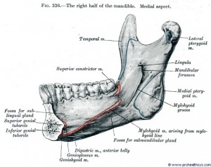
Mylohyoid ridge reduction

© http://www.prohealthsys.com

Mylohyoid ridge is an oblique ridge on the lingual surface of the lower jaw which extends from the level of the roots of the last molar as a bony attachment for the mylohyoid muscles which form the floor of the mouth. Mylohyoid ridge reduction is needed when the gum ridge is sharp and denture pressure can cause significant pain in this area. Continue reading

Preprosthetic Surgery: Preparation of Mouth for Dentures Part 1

© mund-kiefer-gesicht-bremen.de

The prosthetic replacement of lost teeth frequently, though not necessarily, involves surgical preparation of the remaining soft tissues in the mouth. The aim is to leave a satisfactory base for subsequent placement of prosthetic appliances to enable an edentulous or toothless individual to live comfortably with functioning dentures.

Ideally treatment for these procedures should be planned jointly (by a prosthodontist and oral surgeon). However many procedures of preprosthetic surgery are becoming history due to advancements made in dental science such as the introduction of dental implants. Continue reading

Classification of partial dentures Part 2

Criteria 4: Residual Ridge Characteristics

The criteria published for the Classification System for Complete Edentulism are used to categorize any edentulous span present in the partially edentulous patient. Continue reading

Classification of Partial Detures Part 1

PARTIAL DENTURE

Definition

A dental prosthesis that restores one or more but not all of the natural teeth and/or associated parts and that is supported in part by natural teeth, dental implant supported crowns, abutments, or other fixed partial dentures and /or the mucosa; usage: a partial denture should be described as a fixed partial denture or removable partial denture based on the patient’s capability to remove or not remove the prosthesis. Continue reading

Odontogenic myxoma

The odontogenic myxoma is an uncommon benign odontogenic tumor arising from embryonic connective tissue associated with tooth formation. As a myxoma, this tumor consists mainly of spindle shaped cells and scattered collagen fibers distributed through a loose, mucoid material. Continue reading

Calcifying epithelial odontogenic tumor

The calcifying epithelial odontogenic tumor was first described by Pindborg in 1956; hence also called Pindborg’s tumor. The calcifying epithelial odontogenic tumor is a benign odontogenic tumor of epithelial origin that accounts for approximately 1% of all odontogenic tumors. The origin of this neoplasm is not clearly known, although it is generally accepted to be derived from oral epithelium, reduced enamel epithelium, stratum intermedium or dental lamina remnants. It is more common in the posterior part of the mandible of adults in the fourth to fifth decades. There is no gender predilection. It is characterized by squamous epithelial cells, calcifying masses, and homogeneous acellular material admixed with the tumor epithelium and stroma that have been identified as amyloid. Continue reading

Ameloblastoma

Ameloblastoma (from the early English word amel, meaning enamel + the Greek word blastos, meaning germ) is a rare, benign tumor of odontogenic epithelium (ameloblasts, or outside portion, of the teeth during development) much more commonly appearing in the lower jaw than the upper jaw. It was recognized in 1827 by Cusack. This type of odontogenic neoplasm was designated as an adamantinoma in 1885 by the French physician Louis-Charles Malassez. It was finally renamed to the modern name ameloblastoma in 1930 by Ivey and Churchill. Continue reading

Extraction of baby teeth

1. Why extract a baby tooth?

Choosing between extraction and filling or conservation is a very common decision that both dentists and patients have to take every day. With adults the decision making is much easier, if the tooth is restorable and the patient can pay for the treatment then we simply conserve the tooth. If the patient is a child then the process of decision making is much harder as many factors affect the selection of the treatment plan that the dentist have to follow. Continue reading