Le Fort fractures Part 2

Diagnosis

Diagnosis is made based on physical exam findings with confirmation by axial CT. The patient is taken for radiography of the head and neck then after obvious fracture signs the patient is taken to CT scan for more specific anatomic information. To qualify for LeFort Fractures the pterygoid plates must be involved. These are seen posterior to the maxillary sinuses on axial CT and inferior to the orbital rim on coronal slices. Also, the palate is usually mobile on physical exam. Continue reading
Le Fort fractures Part 1

Le Fort fractures (also spelled as LeFort fractures) are types of facial fractures involving the maxillary bone and surrounding structures in a usually bilateral and either horizontal, pyramidal or transverse way. LeFort fractures are classic in facial trauma. The Le Fort fracture was named after French surgeon René Le Fort (1869–1951), who described them in the early 20th century. Continue reading

What Happens at an Appointment with an Endodontist? Part 2

Continued from Part 1

© agd.org

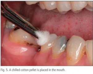
Cold testing

A cold stimulus such as refrigerant spray is sprayed on a cotton pledget and applied to the tooth under investigation.

Heat testing

A heated source such as a GP stick is applied to the tooth. Continue reading

What Happens at an Appointment with an Endodontist? Part 1

© nurhakim.com

Your dentist referred you to an endodontist for a root canal treatment that he/she finds difficult to do. However you started having the jitters about your endodontic appointment since this is a specialist you are seeking. This article will give you a gist on what happens at an appointment with an endodontist. Continue reading

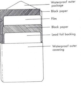
Know More About Xray Films

X-RAY FILMS


Introduction
X-ray film is an image receptor.
It is a type of photographic film, in which an image is formed by
the exit radiation exposing the film.The film is composed of 2
principal components namely the base& emulsion.
Base:
0.2 mm thick & made up of clear, transparent cellulose acetate or
Cellulose nitrate.Cellulose acetate is used because it is less
inflammable. Recently polyesters [polyethylene teraphthalate] are
used.The base should be flexible for easy handling{manipulation}.
Emulsion:Consists of homogenous mixture of gelatin & silver
halide crystals.The gelatin is made from cattle bone.The gelatin is
clear so that it will transmit light & sufficiently porous to allow the
processing chemicals to penetrate it & reaches the silver halide crystals fast without affecting the strength.

Continue reading

Xray Production Part 3

Bremsstrahlung radiation

In an X-ray tube the electrons emitted from the cathode are accelerated towards the metal target anode by an accelerating voltage of typically 50 kV.
The high energy electrons interact with the atoms in the metal target.
Sometimes the electron comes very close to a nucleus in the target and is deviated by the electromagnetic interaction.

Continue reading

Cheek swelling

Cheek pain, at times, tends to be like one of those unwanted guests who arrive without warning, suggest meekly that they’ll be staying only for a couple of days, overstay, and throughout the entire episode, cause immense trouble and discomfort. Cheek pain causes can range from the medically-driven to the hormone-driven (the latter I will illustrate towards the end of this article). Regarding the former, here are some of the possible reasons for cheek pain and swelling. Continue reading

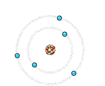
Xray Production Part 1

Introduction
Generation, emission & absorption of radiation occurs at the subatomic level.
Nucleus- two particles- protons & neutrons [nucleons].
Particles that orbit the nucleus are the electrons.
Interest to diagnostic radiology- electrons, protons & neutrons.
number of protons- nucleus of atom- atomic number [Z].
total number of protons & neutrons – nucleus of an atom- mass number [A].

Continue reading

Inferior alveolar nerve Part 2

  • for determining the location of the inferior alveolar nerve in all three dimensions of the mandible (lower jaw). With the software that is available to the dentist, the radiographic information from this study can be used to manipulate the jaw in 360° and provide the most accurate information to the dentist for treatment planning for the placement of the implant or for a third molar extraction. When screening radiographs demonstrate the increased risks for implant placement or third molar extraction, it is below the standard of care for the dentist or oral surgeon to fail to explain the
    advantages of the CT or CBCT as a diagnostic option to the patient before the patient is subjected to the increased risk of nerve injury. Continue reading